Project Description:
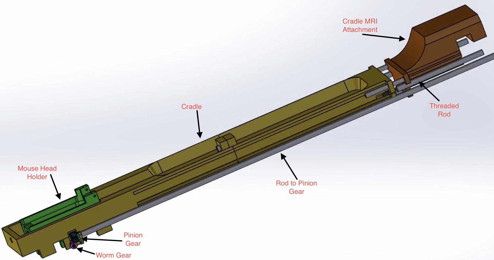
Small Animal Magnetic Resonance Imaging (SA-MRI) is used for preclinical and molecular MRI for small animal in-vivo imaging. SA-MRI combines functional MRI and tractography tensor imaging. Among the challenges of automating fMRI and STI processes is data acquisition as a function of subject positioning. Mismatch in isocentric positioning and inconsistency in subject loading can lead to spatial discrepancies and signal variability. The readjustment process is time-consuming and can lead to inaccuracies in imaging due to manual positioning. A semi-automatic apparatus tailored for aligning subjects within the MRI system proves to be effective in minimizing spatial discrepancies between subject and reference images along with dependence on registration softwares that align the images virtually. The redesigned cradle implements a head holder fashioned along a pair of rods that allow for sliding actuation, in the x-direction, by way of a stepper motor attached to a worm and pinion gear. The back of the cradle is supported by three rods and two screws attached to stepper motors, allowing for the rotation of the screws to move into/out of the machine, z-direction. These changes ensure minimal pain and discomfort for the small animal by using micrometric neck movement. Implementation of the new device will have a positive impact on reducing required anesthesia time and technician fatigue along with the need for manual manipulation of the scanned images.